Why X-Ray Detectable Surgical Sponges Matter in the Operating Room
Every operating room team knows the discipline of the count. Sponges in, sponges out—counted before the procedure, counted before closing,

Managing post-tonsillectomy bleeding and pain remains one of the most clinically significant challenges in outpatient ENT surgery. This review examines current hemostatic strategies, opioid-sparing protocols, and the clinical evidence supporting carboxymethylcellulose-based wound dressings in tonsillectomy and adenoidectomy procedures.
Clinical information notice: This article is intended for licensed healthcare professionals including ENT surgeons, otolaryngologists, surgical center staff, and medical procurement teams. It is provided for informational and educational purposes and does not constitute medical advice. Clinical decisions should be made in accordance with current evidence-based guidelines and individual patient assessment.
Tonsillectomy with or without adenoidectomy (T&A) remains one of the most commonly performed surgical procedures in the United States, with approximately 530,000 procedures performed annually in patients under 15 years of age. Despite advances in surgical technique, post-tonsillectomy hemorrhage (PTH) and post-operative pain continue to represent the most clinically significant complications of the procedure.
Post-tonsillectomy hemorrhage is classified as either primary — occurring within 24 hours of surgery — or secondary — occurring between 24 hours and 10 days post-operatively. Secondary hemorrhage, which typically peaks between days 5 and 10 as eschar separates from the tonsillar fossa, represents the most clinically challenging presentation, with rates reported between 0.5% and 5% depending on surgical technique and patient population.
The intersection of post-operative pain management and hemostasis has become increasingly important as the ENT surgical community responds to national pressure to reduce opioid prescribing in pediatric patients following T&A. Strategies that simultaneously support wound healing, reduce inflammation, and control bleeding are of particular clinical interest.
| Stat | What it means | Source |
|---|---|---|
| 530K | Tonsillectomies performed annually in pediatric patients in the U.S. | American Academy of Otolaryngology–Head and Neck Surgery |
| 7–10 | Peak days for post-operative pain following tonsillectomy | Clinical consensus, AAO-HNS guidelines |
| 0.5–5% | Rate of post-tonsillectomy secondary hemorrhage by technique | Cochrane systematic review, 2020 |
The selection of hemostatic technique in tonsillectomy has evolved considerably over the past two decades. The transition from cold dissection to electrosurgical and coblation techniques has altered bleeding profiles — reducing intraoperative hemorrhage while in some analyses increasing secondary hemorrhage rates due to deeper thermal injury and delayed eschar separation.
Standard hemostatic approaches in tonsillectomy include bipolar electrocautery, harmonic scalpel, coblation, and suture ligation. Each carries distinct bleeding and wound-healing profiles:
| Technique | Intraoperative bleeding | Secondary hemorrhage risk | Wound healing profile |
|---|---|---|---|
| Cold steel dissection | Higher | Lower | Cleaner tissue planes, faster mucosal recovery |
| Bipolar electrocautery | Lower | Moderate | Thermal injury to adjacent tissue; slower healing |
| Coblation | Lower | Moderate–higher | Reduced thermal spread; deeper tissue effects debated |
| Harmonic scalpel | Lower | Moderate | Vibration-based; reduced lateral thermal damage |
Regardless of technique, the post-operative wound bed requires a supportive healing environment. The tonsillar fossa, exposed to the oral microbiome and repeated mechanical stress from swallowing, is particularly vulnerable to secondary hemorrhage during the eschar maturation phase. Hemostatic dressings that support this phase through bioactive wound management represent an emerging adjunct to standard surgical technique.
The post-tonsillectomy wound bed presents a unique biological environment. Following dissection, the exposed tonsillar fossa undergoes a predictable inflammatory and proliferative healing sequence, during which fibrin deposition, neutrophil infiltration, and early angiogenesis occur within the first 48 to 72 hours. The quality of this early healing phase directly influences both secondary hemorrhage risk and pain intensity during the peak recovery window of days 7 through 10 post-operatively.
Carboxymethylcellulose (CMC)-based hemostatic matrices — including etherified variants designed for ENT applications — act through multiple mechanisms to support this healing environment:
Mechanism of action — etherified CMC matrices in ENT wound beds: Etherified carboxymethylcellulose represents an advanced formulation of standard CMC. The etherification process enhances water solubility, pH neutrality (approximately 7.2), and binding affinity to wound surfaces, making it particularly suited to the moist, mechanically active environment of the oropharynx. Unlike oxidized cellulose products, etherified CMC contains no animal, human, or added chemical components, making it appropriate for use in patients with sensitivities or contraindications to animal-derived hemostatic agents.
A prospective clinical trial evaluating the therapeutic effects of Sealk™ on post-tonsillectomy wound sites — conducted with 50 patients (Bunting, E., Hawley, B., 2023) — demonstrated statistically significant improvements in post-operative pain scores on the treated side compared to control.
These findings are consistent with the proposed mechanism of CMC-mediated cytokine modulation and moist wound maintenance. Critically, pain relief was observed to be consistent across the full post-operative recovery window (Days 1–10), including the peak pain period of Days 7–10, which has historically been the most challenging phase for opioid-sparing pain management.
| Result | Detail | Source |
|---|---|---|
| 64% | Of post-tonsillectomy patients reported less pain on the Sealk™-treated side | Bunting, E., Hawley, B. Clinical Trial, 50 pts. 2023 |
| >40% | Average reduction in pain scores across Days 1–10 post-operatively | Bunting, E., Hawley, B. Clinical Trial, 50 pts. 2023 |
“I am no longer prescribing narcotics for pediatric T&A after implementing Sealk. It works so well that patients and parents are requesting it now.” — Dr. K. Smothers, ENT
This testimonial represents the independent opinion and clinical experience of the physician. It should not be interpreted as an FDA-cleared claim or as a representative of all clinical outcomes.
The opioid-sparing imperative in pediatric ENT surgery has intensified following the 2019 FDA boxed warning restricting codeine and tramadol use in children under 12 years of age following tonsillectomy. ENT surgeons and ambulatory surgery centers are under increasing clinical and regulatory pressure to develop effective multimodal analgesia pathways that eliminate opioid dependence for routine T&A recovery.
Current evidence-based approaches to opioid-free or opioid-minimizing T&A pain management incorporate several modalities:
The integration of wound-level hemostatic support into a multimodal analgesia protocol addresses a gap that pharmacological approaches alone cannot fully close: the local inflammatory environment of the healing tonsillar fossa. By reducing cytokine burden at the wound site, CMC-based dressings may attenuate the nociceptive signaling that drives pain intensity during Days 7–10, the period in which opioid prescribing has historically been most difficult to eliminate.
CMC-based hemostatic matrices designed for ENT applications are applied following standard tonsillar dissection and primary hemostasis. The four-step application protocol is straightforward and adds minimal time to the operative procedure:
The translucent gel formed on irrigation allows intraoperative wound monitoring without requiring dressing removal. As a water-soluble material, the matrix is fully resorbed and leaves no residue — eliminating the secondary hemorrhage risk associated with mechanical dressing removal.
Key characteristics — CMC hemostatic matrices for ENT:
The following images were taken intraoperatively by Dr. Jonathan Simmonds, ENT, during a pediatric tonsillectomy in which Sealk™ was applied to the tonsillar fossa. The patient’s family has consented to the use of these images for clinical and educational purposes.
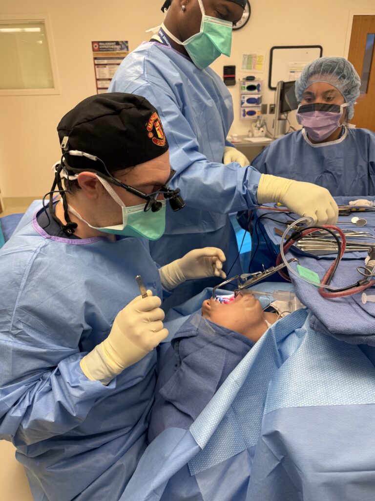
Dr. Jonathan Simmonds (front left) and surgical team in the operating room during the procedure.
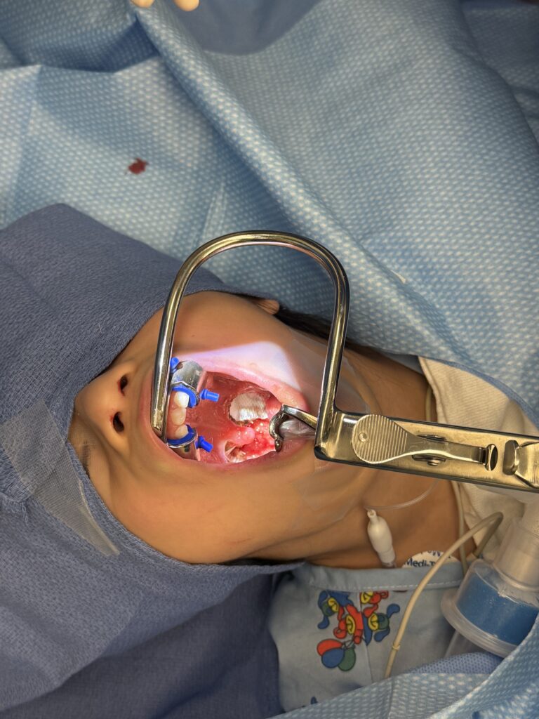
Intraoral view with mouth gag retraction, showing Sealk™ placed in the tonsillar fossa following dissection.
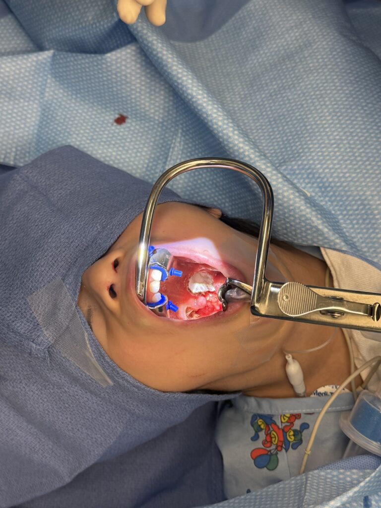
Sealk™ matrix conforming to the tonsillar fossa wound bed, with the contralateral fossa visible.
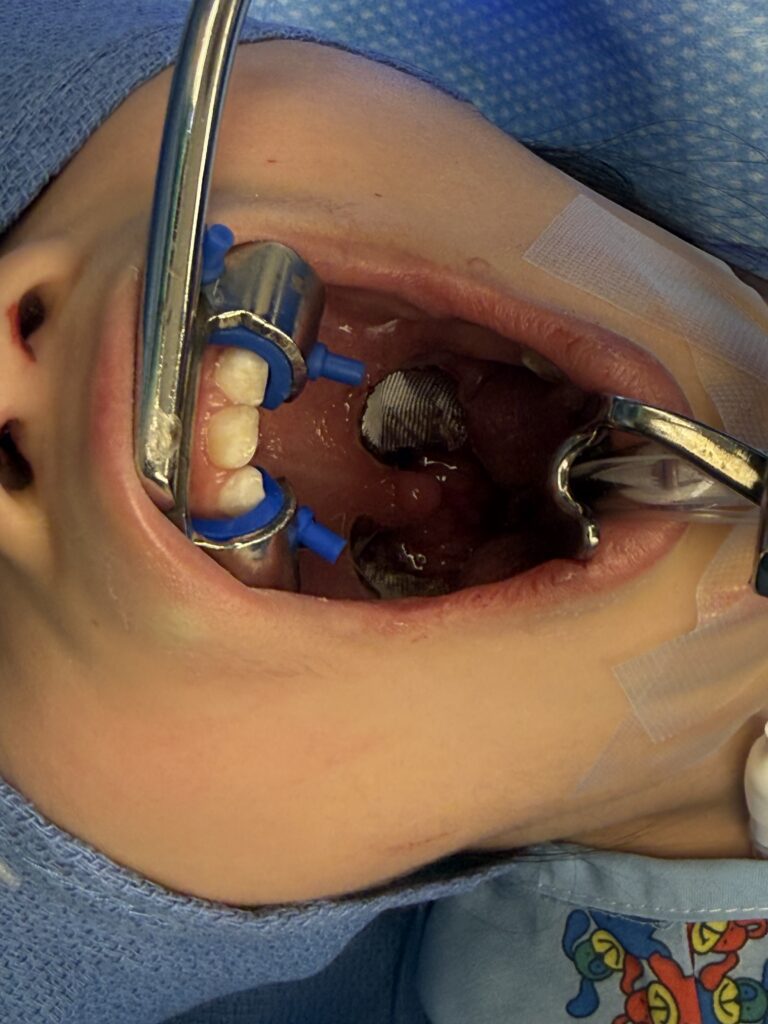
Close-up intraoral view showing the Sealk™ dressing adhered to the wound surface and active hemostasis in the adjacent fossa.
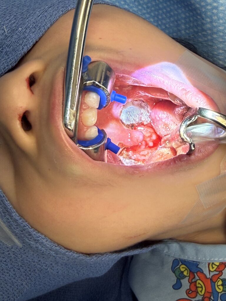
Close-up view following placement, with the Sealk™ matrix visible against the tonsillar bed.
Surgeon testimonial in development: Dr. Jonathan Simmonds has agreed to provide a video testimonial discussing his experience — and the patient’s experience — with Sealk™ in this case. Details and footage to follow.
The clinical and operational case for adopting bioactive hemostatic dressings in the ASC setting extends beyond individual patient outcomes. For ambulatory surgery centers managing high volumes of T&A procedures, the ability to demonstrate meaningful reductions in post-operative pain, opioid prescribing rates, and potentially secondary hemorrhage-related return visits represents a significant quality metric.
ASC quality reporting programs increasingly capture post-discharge pain scores, opioid prescribing rates, and unplanned return visits — all of which can be favorably influenced by systematic wound bed management as part of the post-operative protocol. Procurement teams evaluating hemostatic supplies for T&A programs should assess products not only on unit cost but on total clinical value, including potential reductions in post-operative complications and opioid-related outcomes reporting.
For ENT-focused surgery centers, the availability of a dedicated, FDA-cleared ENT hemostatic dressing from a specialty distributor with standing inventory and responsive service represents a meaningful operational advantage over general surgical supply channels, particularly during periods of supply constraint.
Post-tonsillectomy hemostasis and pain management represent interconnected clinical challenges in outpatient ENT surgery. Carboxymethylcellulose-based hemostatic dressings address both through bioactive wound support — providing mechanical hemostasis, activating tissue regeneration pathways, reducing inflammatory cytokine burden, and maintaining the moist wound environment associated with improved mucosal healing and reduced pain intensity.
Clinical trial data from a 50-patient prospective study demonstrates a 64% rate of patient-reported pain reduction on the treated side and greater than 40% average pain reduction across the full post-operative recovery period. These findings support the integration of CMC-based hemostatic wound dressings into opioid-sparing T&A protocols as a complement to standard pharmacological multimodal analgesia.
For ENT surgeons and surgical center procurement teams seeking to reduce opioid prescribing in pediatric T&A populations while supporting optimal wound healing, etherified carboxymethylcellulose hemostatic matrices represent a clinically meaningful, operationally straightforward adjunct to current practice.
Distributed exclusively by Thomas Jane Medical Sealk™ Advanced Hemostatic Dressing FDA-cleared · ENT · Plant-based · BloodSTOP® iX · Available for surgical center evaluation
© 2026 Thomas Jane Medical, LLC · 1356 N. Emporia Ave, Wichita, KS 67214 · 316-251-0920 · purchase@thomasjanemedical.com